There is growing evidence that depression can reduce gray matter volume in the following regions:
Hippocampus: This part of the brain is important for learning and memory, The Hippocampus connects to other parts of the brain that control emotions and respond to stress hormones:
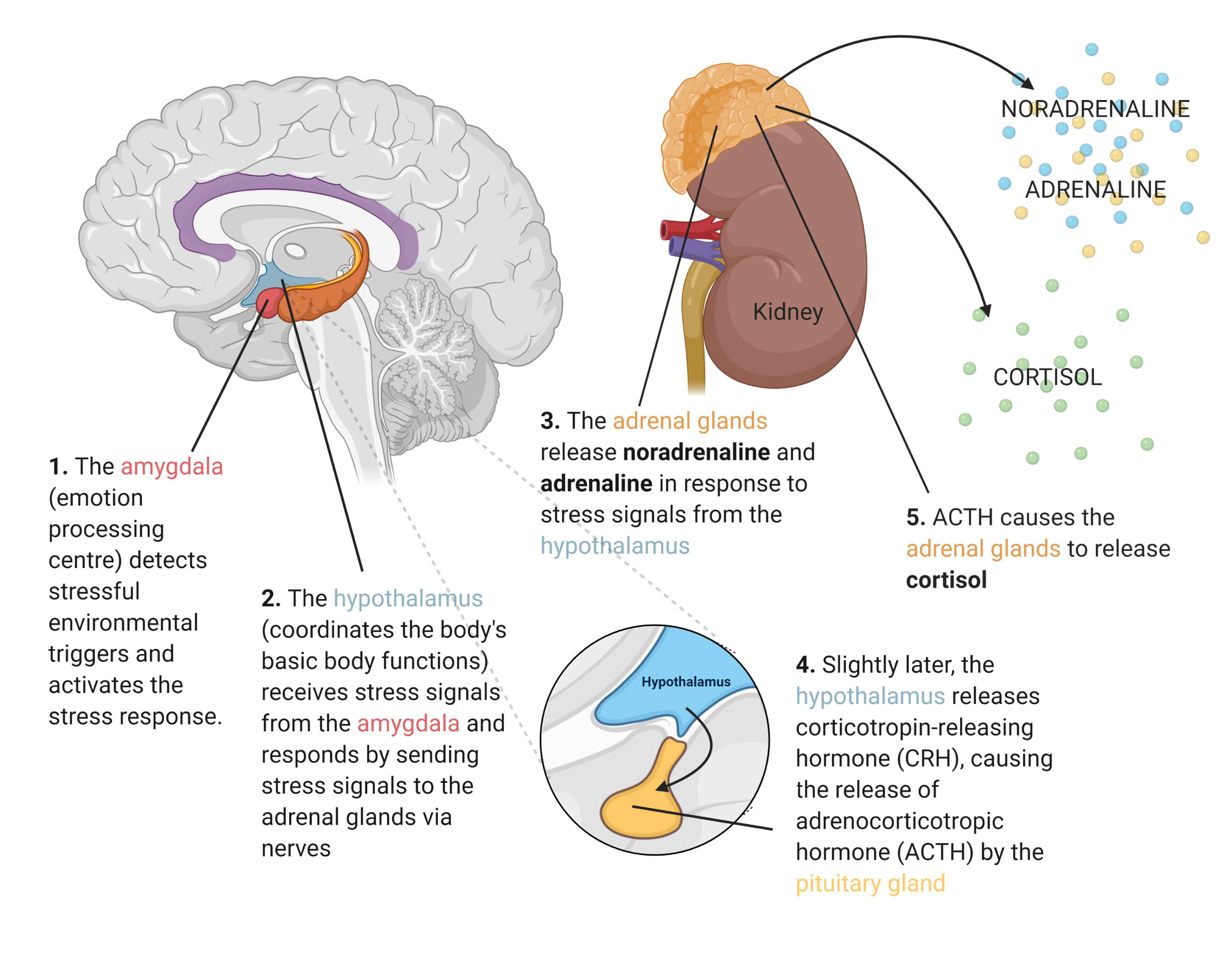
1. Amygdala (emotional processing center): detects environmental stressors and activates the stress response.
2. Hypothalamus (coordinates basic body functions): receives stress signals from the amygdala and responds by sending stress signals to the adrenal glands via nerves.
3. The adrenal glands release noradrenaline and adrenaline in response to stress signals from the hypothalamus.
4. The hypothalamus then secretes corticotropin-releasing hormone (CRH), which causes the pituitary gland to secrete adrenocorticotropic hormone (ACTH).
5. ACTH causes the adrenal glands to secrete cortisol
Prefrontal cortex: This area plays a role in thinking and planning.
Other brain regions that shrink in depression include the thalamus, caudate nucleus, and insula.

Figure 1: Brain Regions Involved in Stress Hormones
Studies show that stress and depression can increase gray matter volume loss. The more severe the depression, the greater the gray matter volume loss. When these areas don’t work properly, you may have problems like:
In situations of chronic stress, excessive glucocorticoid release can eventually cause hippocampal atrophy. Because the hippocampus inhibits the HPA axis, atrophy may lead to chronic activation of the HPA axis, which may increase the risk of developing disease. Because the HPA axis is central to stress processing. Mechanisms being examined include antagonism of the glucocorticoid receptor, corticotropin-releasing factor 1 (CRF-1) receptor, and vasopressin 1B receptor.

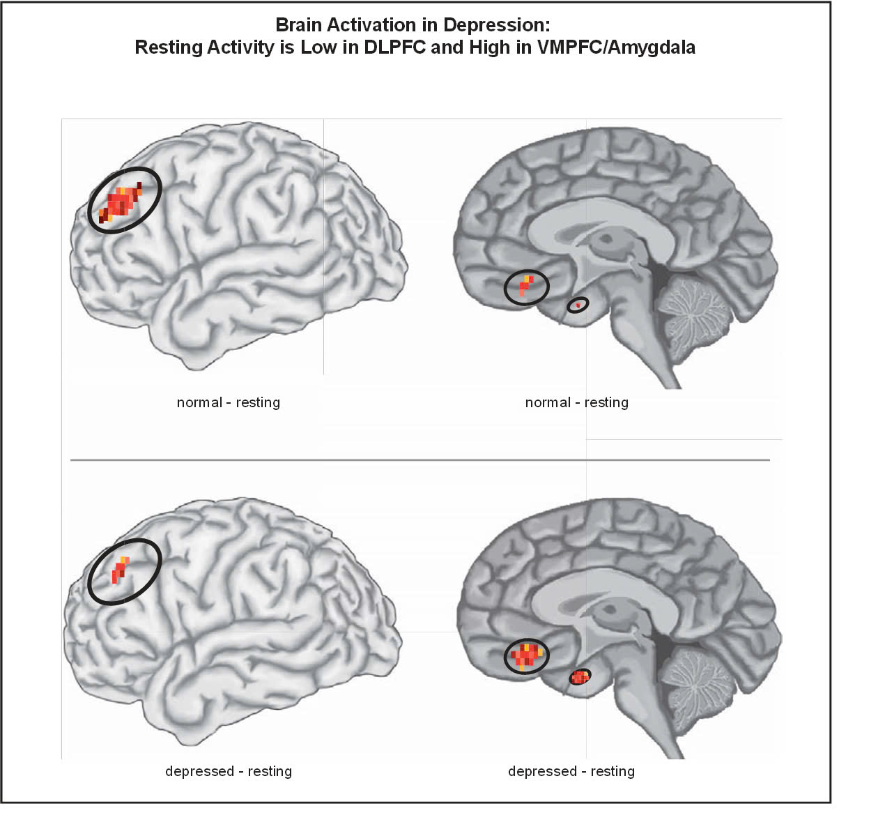
Figure 2: Brain Activity in Depression
The more severe the depression, the greater the loss of gray matter volume. Neuroimaging studies of brain activation show that resting activity in the dorsolateral prefrontal cortex (DLPFC) of depressed patients is low compared with activity in non-depressed individual, while resting activity in the amygdala and ventromedial prefrontal cortex (VMPFC) of depressed patients is higher than that of non-depressed individuals.
To minimize damage to the brain, you should see a doctor and get treatment early, following the instructions of a psychiatrist. This will ensure your ability to work, study, and quality of life.
Reference: Stephen M. Stahl “Neuroscientific Basis and Practical Application”, Stahl’s Essential Psychopharmacology. Fourth Edition. P 349 – 354.